Home

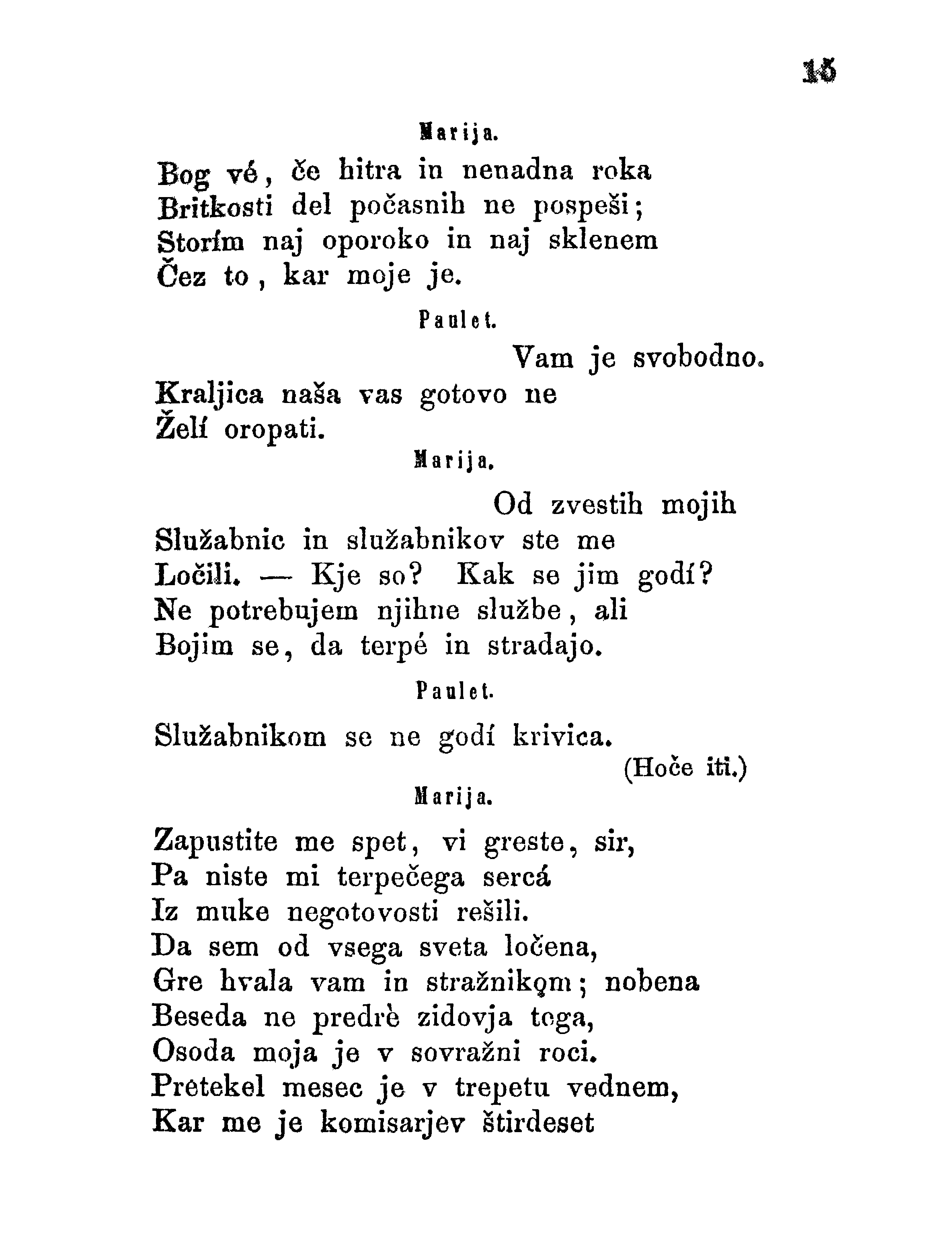
apvalkalas studija kolonija sem vam očitno pisala prenaglo Miegamasis Kulnas Pasviręs brūkšnys

Transmission electron microscopy (TEM) of developed oocytes (IV–V) of... |  Download Scientific Diagram
Transmission electron microscopy (TEM) of developed oocytes (IV–V) of... | Download Scientific Diagram

Scanning electron microscopy (SEM) before (left) and after (right)... |  Download Scientific Diagram
Scanning electron microscopy (SEM) before (left) and after (right)... | Download Scientific Diagram

SEM images for cellulose acetate membranes:A )A symmetrical B)... |  Download Scientific Diagram
SEM images for cellulose acetate membranes:A )A symmetrical B)... | Download Scientific Diagram

A) SEM images of platelets and erythrocytes adhesion on the surface of... |  Download Scientific Diagram
A) SEM images of platelets and erythrocytes adhesion on the surface of... | Download Scientific Diagram

SEM analysis of external platelet morphology. External morphology of... |  Download Scientific Diagram
SEM analysis of external platelet morphology. External morphology of... | Download Scientific Diagram

SEM images for the adhered platelets on the inner surface of the hollow...  | Download Scientific Diagram
SEM images for the adhered platelets on the inner surface of the hollow... | Download Scientific Diagram

Scanning electron microscopy (SEM) before (left) and after (right)... |  Download Scientific Diagram
Scanning electron microscopy (SEM) before (left) and after (right)... | Download Scientific Diagram

SEM, TEM images and electron diffraction patterns of (a) precipitated... |  Download Scientific Diagram
SEM, TEM images and electron diffraction patterns of (a) precipitated... | Download Scientific Diagram

SEM images of blank polycarbonate membrane before and after growth of... |  Download Scientific Diagram
SEM images of blank polycarbonate membrane before and after growth of... | Download Scientific Diagram

je na razpolago pod dovoljenjem Creative

SEM images of the alginate fibers' cross-section with different CN... |  Download Scientific Diagram
SEM images of the alginate fibers' cross-section with different CN... | Download Scientific Diagram

SEM image of conventional platelets (A) and PCT platelets (B) show that...  | Download Scientific Diagram
SEM image of conventional platelets (A) and PCT platelets (B) show that... | Download Scientific Diagram

Methods: TEM and SEM The morphology of the nanoparticles was examined using  a transmission electron microscopy (TEM) at UCL, Sch
Methods: TEM and SEM The morphology of the nanoparticles was examined using a transmission electron microscopy (TEM) at UCL, Sch

SEM images of platelet adhesion on different surfaces (A)... | Download  Scientific Diagram
SEM images of platelet adhesion on different surfaces (A)... | Download Scientific Diagram

Scanning electron microscopy (SEM) before (left) and after (right)... |  Download Scientific Diagram
Scanning electron microscopy (SEM) before (left) and after (right)... | Download Scientific Diagram

Scanning electron microscopy (SEM) micrographs of the alginate/gelatin... |  Download Scientific Diagram
Scanning electron microscopy (SEM) micrographs of the alginate/gelatin... | Download Scientific Diagram

Diagnostic Electron Microscopy of Viruses With Low-voltage Electron  Microscopes
Diagnostic Electron Microscopy of Viruses With Low-voltage Electron Microscopes

A SEM view of the apical region of Euglena, after the chitosan... |  Download Scientific Diagram
A SEM view of the apical region of Euglena, after the chitosan... | Download Scientific Diagram

Methods: TEM and SEM The morphology of the nanoparticles was examined using  a transmission electron microscopy (TEM) at UCL, Sch
Methods: TEM and SEM The morphology of the nanoparticles was examined using a transmission electron microscopy (TEM) at UCL, Sch

Diagnostic Electron Microscopy of Viruses With Low-voltage Electron  Microscopes
Diagnostic Electron Microscopy of Viruses With Low-voltage Electron Microscopes

Language Server, Jožef Stefan Institute FPG_00061-

Platelet morphology. SEM images of platelets adhered onto... | Download  Scientific Diagram
Platelet morphology. SEM images of platelets adhered onto... | Download Scientific Diagram

Diagnostic Electron Microscopy of Viruses With Low-voltage Electron  Microscopes
Diagnostic Electron Microscopy of Viruses With Low-voltage Electron Microscopes

SEM image showing attachment and biofilm formation by Shigella boydii... |  Download Scientific Diagram
SEM image showing attachment and biofilm formation by Shigella boydii... | Download Scientific Diagram

Amoeba sem hi-res stock photography and images - Alamy

A SEM view of the apical region of Euglena, after the chitosan... |  Download Scientific Diagram
A SEM view of the apical region of Euglena, after the chitosan... | Download Scientific Diagram

Scanning electron microscopy (SEM) micrographs of the alginate/gelatin... |  Download Scientific Diagram
Scanning electron microscopy (SEM) micrographs of the alginate/gelatin... | Download Scientific Diagram